What X-Rays Tell Us About Spinal Structure And Function
X-rays were first discovered by William Roentgen in 1895, coincidentally the same year that the discoverer of chiropractic, D.D. Palmer established the chiropractic profession with the first adjustment. D.D.'s son, B.J., brought X-ray analysis into the profession with the first X-ray machine in 1910. So, X-rays have always been a part of chiropractic analysis in some manner.
Like many technological advancements, X-rays have undergone a remarkable evolution. In the early days, exposures were prolonged, and safety measures were inadequate, leading to adverse reactions to radiation among doctors and technicians. Today, X-rays have evolved to a point where they bear no resemblance to their early counterparts, with enhanced safety and efficiency.
When I started my first chiropractic practice in 1973, I had an x-ray machine. I also had a darkroom to develop the films. We had a 'safe' light to see in the dark. The films were taken from cassettes and placed on hangers in the tanks to bring out the image. The next step was an automatic processing machine in which the films were again taken out of cassettes in the dark and fed through the processor. From there, digital X-rays came about, no longer necessitating a darkroom, but we still used cassettes. The last X-ray processing I used was digital X-ray without the need for cassettes; the images appeared directly on a computer screen quickly.
X-rays reveal obvious pathologies. Several times, I picked up fractures and anomalies that I couldn't have known about without the images. In addition to pathology, such as loss of bone density, X-rays can help detect degenerative processes, which often include overgrowth of bone that can directly impinge on nerves.
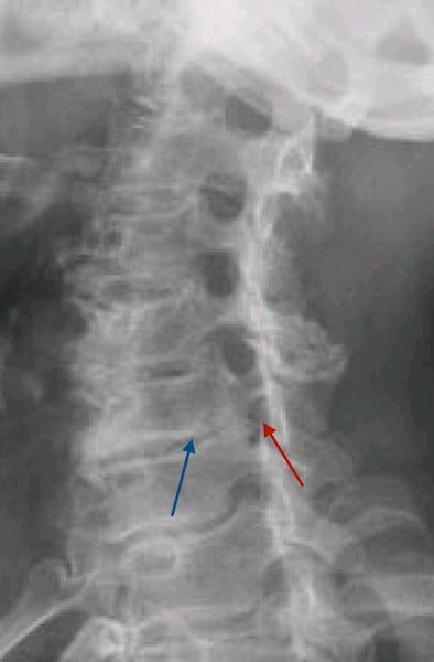
In the image below, the red arrow points to the nerve opening, which has been severely compromised compared to the ones above. The poor nerve is trapped. This process has occurred because of the severe degeneration between the two vertebrae shown by the blue arrow. The opening below has also been reduced in size.
While chiropractors recognize these pathologies, other structural and functional characteristics visible on X-rays assist in spinal analysis.
Keep reading with a 7-day free trial
Subscribe to The Truth Patrol to keep reading this post and get 7 days of free access to the full post archives.


